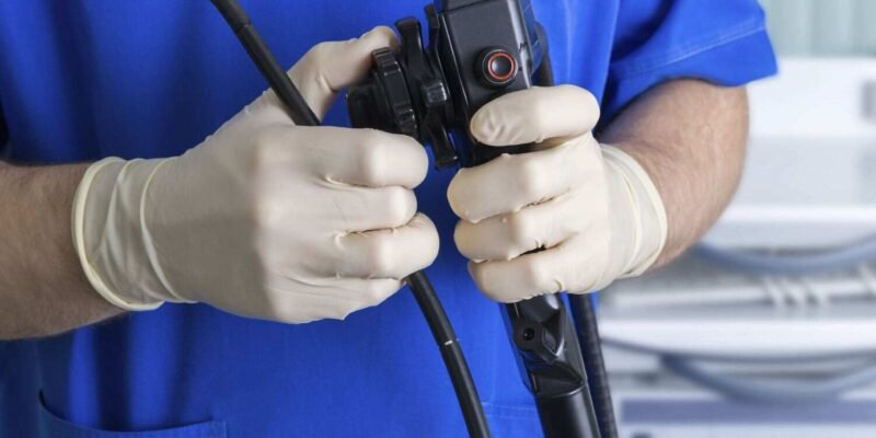
Endoscopes are essential medical instruments that allow healthcare professionals to examine the internal structures of a patient’s body with minimal invasion. They have revolutionized diagnostics and treatments by enabling doctors to visualize organs, tissues, and other bodily features without requiring major surgical procedures. The effectiveness of an endoscope depends on its parts working in harmony. In this article, we will explore the essential parts of an endoscope and their respective functions.
Understanding the Basic Components of an Endoscope
An endoscope consists of several integral parts, each serving a specific function to ensure the device’s successful operation. The endoscope’s components must be precise and reliable, whether used for diagnostic purposes, such as identifying cancer, or for procedures like biopsies and removing foreign objects.
The Insertion Tube
The insertion tube is one of the most crucial components of the endoscope. This long, flexible tube is inserted into the body, allowing the device to navigate different anatomical areas. The insertion tube is designed for both durability and flexibility, enabling it to maneuver through tight spaces without causing harm to the body. It also contains various channels for light, imaging, and medical tools such as forceps or biopsy needles.
The insertion tube’s flexibility is essential for doctors to move the endoscope through complex bodily structures, such as the gastrointestinal tract or the respiratory system. The robustness of this part of the endoscope ensures that it can withstand the movements and forces encountered during the procedure.
Camera and Lens
The camera and lens are the endoscope’s “eyes.” Positioned at the tip of the insertion tube, the camera captures real-time images and transmits them to a monitor for the healthcare provider to analyze. Modern endoscopes often use high-definition cameras, which provide clear and detailed pictures of internal body parts.
The lens is specially designed to focus light and enhance the clarity of the images captured by the camera. This essential part is necessary for doctors to observe abnormalities in internal organs, such as tumors or foreign objects. The quality of the camera and lens directly influences the accuracy of the diagnosis and treatment.
Light Source
The light source provides the necessary illumination for the endoscope to function effectively. Positioned within the insertion tube, it uses fiber optic technology to shine light on the examined area. Clear visibility is crucial for a successful endoscopic procedure, as it allows the healthcare provider to see the details within the body, even in dark or hard-to-reach areas.
Endoscopes typically use a powerful light source to enhance image quality, ensuring doctors have a clear view of the examined organs or tissues. Even the best camera would only provide valuable images with proper lighting.
Biopsy Channel
The biopsy channel is a small but vital part of the endoscope. It allows for the insertion of medical instruments, such as biopsy forceps, which are used to collect tissue samples for further analysis. The biopsy channel allows doctors to perform diagnostic procedures, such as detecting cancer or other abnormalities, without the need for invasive surgery.
This component is critical for procedures like colonoscopies, where tissue samples may be needed to identify signs of disease. The biopsy channel facilitates the collection of these samples minimally invasively, reducing the risk of complications and speeding up the recovery process.
Suction Channel
The suction channel removes fluids, debris, or mucus from the examined area. In many cases, bodily fluids can obstruct the view, making it difficult for doctors to assess the location accurately. The suction channel allows healthcare providers to clear the workspace, ensuring the camera has an unobstructed view of the observed organs or tissues.
For example, in procedures such as bronchoscopy or colonoscopy, the suction channel helps remove excess fluids or mucus, keeping the area clear for a more accurate examination. It’s essential to ensure that the endoscope functions effectively during procedures.
Control Mechanism
The endoscope’s control mechanism includes buttons, levers, or a joystick that allows the healthcare provider to guide the device during the procedure. This part is crucial for ensuring that the endoscope is maneuvered precisely, allowing it to navigate through complex anatomical structures easily.
The control mechanism allows the healthcare provider to adjust the camera’s angle, regulate the light intensity, and operate other endoscope features. This ensures the procedure is performed efficiently and accurately, with minimal discomfort to the patient.
Outer Sheath
The outer sheath is a protective covering that encases the insertion tube and its internal components. It protects the delicate parts of the endoscope from external damage while also providing a sterile barrier to prevent infection. The outer sheath must be durable yet flexible enough to allow movement during the procedure.
In some cases, disposable outer sheaths are used to reduce the risk of cross-contamination between patients further. This ensures that each procedure is conducted safely, minimizing the risk of infection during diagnostic or therapeutic procedures.
Display Monitor
The display monitor is where the images captured by the camera are displayed in real time for the healthcare provider to analyze. The monitor allows the doctor to closely observe the area being examined and make informed decisions about the next steps in the procedure.
Modern endoscopes often use high-resolution monitors that display images in great detail, allowing doctors to identify even the slightest abnormalities. The clarity and size of the monitor are essential for accurate diagnosis and treatment planning.
Why Endoscope Parts Matter for Medical Procedures
Each part of an endoscope plays an essential role in ensuring the success of a medical procedure. Without any one of these components, the device would not be able to perform its intended functions. The camera’s precision, the insertion tube’s flexibility, and the light source’s brightness all work together to make endoscopy an invaluable tool for modern medicine.
Whether it’s for routine screenings like colonoscopies or more complex procedures like arthroscopies, the quality and reliability of endoscope parts directly impact the procedure’s success and the patient’s safety.
Conclusion
Endoscopes are remarkable tools that have transformed modern medicine. They allow healthcare professionals to perform diagnostic and therapeutic procedures with minimal invasion. The various parts of an endoscope, such as the insertion tube, camera, light source, and biopsy channel, are all designed to work together seamlessly. Understanding these essential parts helps emphasize the importance of high-quality endoscopes in delivering accurate, efficient, and safe medical care.
Just like a natural cotton dress offers comfort and adaptability in daily life, endoscope parts must provide precision, flexibility, and reliability to ensure effective procedures. These components allow healthcare providers to perform detailed examinations and make life-saving diagnoses while minimizing the risk to patients.
Endoscopic technology continues to evolve, along with the parts that make these medical devices possible. As advancements continue, the future of endoscopy will offer even greater accuracy and ease of use, enhancing patient outcomes and improving healthcare delivery worldwide.
Also read interesting articles at Disboard.co.uk


